Publikationen & Bücher
Publikationen - Merz E. Can prenatal testing in the first trimester be performed whithout ultrasound? Ultraschall Med. 2017;38:126-128
- Merz E, Pashaj S. Advantages of 3D ultrasound in the assessment of of fetal abnormalities. J Perinat Med. 2017;45:643-650
- Merz E, Thode C, Eiben B, Wellek S. Prenatal Risk Calculation (PRC) 3.0: An extended DOE-based first-trimester screening algorithm allowing for early blood sampling. Ultrasound Int Open. 2016;2:E19-26
Merz E, Eichhorn KH, Madjar H, Hackelöer BJ, Degenhardt F.
Indication for and possibilities of gynecological breast sonography after the introduction of
mammography screening in Germany.
Ultraschall Med. 2009;30:3-5
Merz E, Thode C, Alkier A, Eiben B, Hackelöer BJ, Hansmann M, Huesgen G,
Kozlowski P, Pruggmaier M, Wellek S.
A new approach to calculating the risk of chromosomal abnormalities with first-trimester screening data.
Ultraschall Med. 2008;29:639-645 Merz E, Thode C, Wellek S, Alkier A, Eiben B, Hackeloer BJ, Hansmann M, Huesgen G, Kozlowski P, Pruggmaier M.
Fetal Medicine Foundation Germany (FMF-D): a new approach to calculating the risk of chromosomal abnormalities with first-trimester screening data (11+1 to 14+0 weeks).
Ultrasound Obstet Gynecol. 2007;30:542-543 Merz E, Benoit B, Blaas HG, Baba K, Kratochwil A, Nelson T, Pretorius D,
Jurkovic D, Chang FM, Lee A; ISUOG 3D Focus Group.
Standardization of three-dimensional images in obstetrics and gynecology: consensus statement.
Ultrasound Obstet Gynecol. 2007;29:697-703 Merz E The fetal nasal bone in the first trimester - precise assessment using 3D Sonography.
Ultraschall Med. 2005;26:365-366 Merz E, Welter C.
2D and 3D Ultrasound in the evaluation of normal and abnormal fetal anatomy in the second and third trimesters in a level III center.
Ultraschall Med. 2005;26:9-16 Merz E, Meinel K, Bald R, Bernaschek G, Deutinger J, Eichhorn K, Feige A, Grab D, Hackeloer BJ, Hansmann M,
Kainer F, Schillinger W, Schneider KT, Staudach A, Steiner H, Tercanli S, Terinde R, Wisser J; DEGUM; Fetal Medicine Foundation.
DEGUM Level III recommendation for "follow-up" ultrasound examination (= DEGUM Level II) in the 11 - 14 week period of pregnancy.
Ultraschall Med. 2004;25:299-301 Merz E, Eichhorn KH, Hansmann M, Meinel
Qualitätsanforderungen an die weiterführende differential-diagnostische Ultraschallunter-suchung in der pränatalen Diagnostik (= DEGUM-Stufe II) im Zeitraum 18 bis 22 Schwangerschaftswochen.
Ultraschall Med. 2002;23:11-12 Merz E, Bahlmann F, Welter C, Miric-Tesanic D
Transvaginale 3D-Sonographie in der Frühgravidität.
Gynäkologe 1999;32:213-219 Merz E, Miric-Tesanic D, Bahlmann F, Weber G, Hallermann C
Prenatal sonographic chest and lung measurements for predicting severe pulmonary hypoplasia.
Prenat. Diagn.1999;19: 614-619 Merz E, Weber G, Bahlmann F, Kießlich R
A new sonomorphologic scoring-system (Mainz Score) for the assessment of ovarian tumors using transvaginal ultrasonography.
Ultraschall Med.1998;19: 99-107 Merz E, Weber G, Bahlmann F, Miric-Tesanic D
Application of transvaginal and abdominal 3D-ultrasound for the detection or exclusion of malformations of the fetal face.
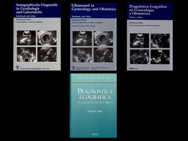
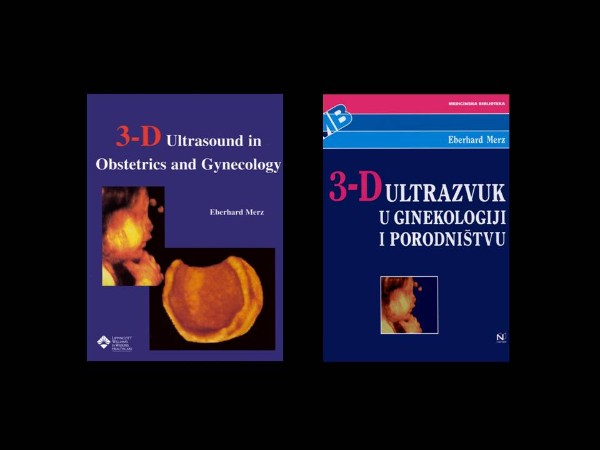
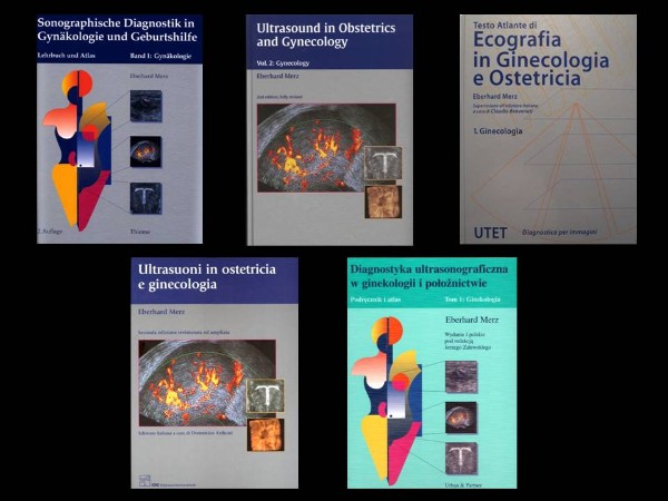
Ultrasound Obstet. Gynecol. 1997;9:237-243 Bücher 


|


